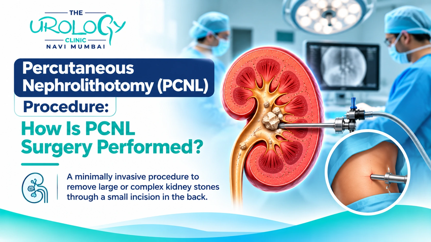


A kidney stone can become a complicated health concern when it grows beyond a size that the body can naturally eliminate. What may initially appear as a small deposit of minerals can gradually develop into a larger formation inside the kidney. When such stones interfere with urine drainage, cause repeated discomfort or increase the risk of infection, surgical treatment may become necessary. The Percutaneous Nephrolithotomy Procedure is one of the established surgical options used in these situations.
Commonly shortened to PCNL, this operation gives the surgeon access to the kidney through a small opening in the patient’s back. Specialised instruments are used to locate, break and extract the stone material. It is particularly useful for patients with substantial stone deposits that are unlikely to be managed effectively through certain non-surgical treatments.
A recommendation for kidney stone surgery can naturally raise several questions. How does the operation work? Is hospital admission necessary? What happens after the stones are removed? This article covers these aspects to help patients become familiar with the treatment process.
The kidneys maintain the body’s fluid and mineral balance while filtering waste products from the blood. Urine carries these waste substances out of the body. When certain minerals become highly concentrated, they can combine and form solid crystals.
Over time, these crystals may join together and develop into kidney stones. Their behaviour depends on their composition, dimensions and location within the urinary system.
A small stone may travel towards the bladder and leave the body during urination. A larger stone may remain inside the kidney, occupy its collecting spaces or obstruct the passage through which urine flows.
This is where treatment planning becomes important. A stone that occupies a large portion of the kidney may require direct removal rather than relying on the body’s natural elimination process. The Percutaneous Nephrolithotomy Procedure is designed to address this type of stone burden.
The decision to recommend PCNL is made after examining the patient’s medical condition and imaging results. Stone size is an important factor, but it is not the only consideration.
Patients with kidney stones measuring more than 2 cm are commonly evaluated for this procedure. It may also be appropriate when several stones are present, particularly if they are distributed across different parts of the kidney.
Another condition that may require this approach is a staghorn calculus. This type of stone develops a branching shape and can extend into multiple sections of the kidney’s collecting system. Its structure can make treatment more complicated.
PCNL may also be considered when previous stone removal treatments have not achieved adequate clearance. The urologist evaluates the remaining stone material and determines whether another procedure is necessary.
The suitability of the Percutaneous Nephrolithotomy Procedure also depends on factors such as kidney anatomy, bleeding risk, infection status and other existing medical conditions.
Preparation begins well before the actual operation. The surgeon needs a clear picture of the kidney’s condition and the patient’s general health to plan the procedure safely.
A CT scan is often useful because it provides detailed images of the stone’s position, size and relationship with surrounding structures. Depending on the clinical situation, ultrasound or other imaging tests may also be requested.
Blood investigations help assess kidney function, haemoglobin levels and other relevant parameters. Urine testing, including a urine culture when indicated, helps identify infections that may need treatment before surgery.
Patients should also share information about their regular medicines, allergies and previous operations. Particular attention is given to blood-thinning medicines because these can influence surgical bleeding risk. Medication adjustments must be made only according to the treating doctor’s instructions.
This assessment helps the surgical team decide the appropriate access route, instruments and postoperative care requirements.
Although the technical details can differ between patients, the operation follows a planned sequence. The following stages describe how the surgeon generally approaches kidney stone removal.
The patient receives general anaesthesia, which keeps them asleep during the operation. The surgical team prepares the required area and positions the patient according to the selected surgical approach.
The surgeon identifies a suitable entry location using imaging guidance. A fine needle is introduced through the skin into the kidney’s collecting system. This initial access is carefully expanded to accommodate the surgical instruments.
The size and direction of the access passage are selected according to the stone location and the patient’s anatomy.
A nephroscope, which is a specialised viewing instrument, is passed through the newly created passage. It allows the surgeon to see the internal kidney structures and identify the stone deposits that require removal.
Large stones cannot always be extracted in one piece. Therefore, the surgeon uses specialised energy-based equipment to divide them into manageable fragments.
Depending on the clinical requirements, laser, ultrasonic or pneumatic devices may be selected. The choice depends on the equipment available, stone characteristics and surgical plan.
Once the stone has been broken down, the fragments are removed through the access tract. The surgeon examines the treated area to assess whether additional stone material needs attention.
After completing the stone removal, the surgeon determines whether a temporary drainage arrangement is required. A nephrostomy tube may be placed through the back, while a ureteral stent may be used internally to support urine flow.
Not every patient requires the same arrangement. The decision depends on the surgical findings and the patient’s recovery needs.
Patients may come across different names when researching kidney stone surgery. Standard PCNL and Mini-PCNL are two variations of the same general surgical approach.
Standard PCNL uses a conventional-sized access tract and instruments. It is commonly selected for substantial or complicated stone deposits where direct access and extraction are required.
Mini-PCNL uses a smaller access passage and narrower instruments. It may be considered for selected stone sizes and locations. The smaller tract can offer certain advantages, including reduced blood loss in some circumstances, although the procedure may require more time.
Neither method is automatically suitable for everyone. The surgeon considers the stone burden, kidney structure and patient’s medical history before selecting an approach.
The period immediately following surgery is mainly focused on observation. The medical team checks the patient’s blood pressure, pulse, urine output and general condition.
Some patients notice a reddish appearance in their urine for a short period. Discomfort around the entry point may also occur. The intensity and duration of these symptoms vary, and the healthcare team assesses them during recovery.
If a nephrostomy tube or ureteral stent has been placed, the patient receives instructions regarding its care and removal. These devices should never be adjusted or removed without medical guidance.
The length of hospital admission depends on how the patient responds to surgery. Discharge is considered when the treating team is satisfied with the patient’s recovery and ongoing care arrangements.
The Percutaneous Nephrolithotomy Procedure is an established surgical treatment, but it is not free from risks. Understanding possible complications helps patients recognise why postoperative monitoring and follow-up are important.
Bleeding may occur because the surgeon creates an access passage into the kidney. In some cases, significant bleeding may require a blood transfusion or another intervention.
Urinary infections are another possible complication. Fever, chills and worsening discomfort after surgery require medical assessment. In uncommon situations, injury to nearby organs or surrounding tissues may occur during access creation.
Some patients may also experience urine leakage or have residual stone fragments after the operation. Depending on their size and location, these fragments may require observation or additional treatment.
The possibility of complications varies with stone complexity, existing infection, kidney anatomy and the patient’s general health.
Returning to everyday activities should happen gradually. The body needs time to recover from anaesthesia and the surgical access created during the operation.
Patients should follow the prescribed medication schedule and maintain fluid intake according to their doctor’s recommendations. People with certain kidney or heart conditions may need individualised fluid advice.
During the initial recovery period, heavy lifting and strenuous exercise should be avoided until medical clearance is provided. Walking and other light activities may be resumed as advised by the treating team.
The incision site should be kept clean according to the wound-care instructions. If a stent has been inserted, its planned removal date should be confirmed before leaving the hospital.
A patient should seek medical attention if they experience high fever, chills, severe pain, inability to pass urine or heavy bleeding. Waiting for such symptoms to disappear without medical assessment is not advisable.
Removing an existing stone does not necessarily stop future stones from forming. The underlying conditions that contributed to mineral accumulation may continue even after successful surgery.
For this reason, stone analysis can be useful after the operation. It identifies the material from which the stone was formed and may help the doctor recommend appropriate preventive measures.
Some patients may need a metabolic evaluation to assess urine composition and identify factors that encourage crystal formation. Depending on the findings, the doctor may suggest dietary changes or specific medication.
Adequate hydration is generally an important part of kidney stone prevention. Reducing excessive salt consumption and following dietary recommendations based on the stone type may also help.
The purpose of follow-up is not only to check the surgical outcome but also to reduce the possibility of another stone developing.
Kidney stones that occupy a large part of the kidney may require a treatment approach that allows direct access and removal. The Percutaneous Nephrolithotomy Procedure provides this access through a small opening in the skin and uses specialised instruments to fragment and extract the stone material.
The treatment journey includes detailed investigations, surgical planning, anaesthesia, stone removal and postoperative observation. Recovery requirements differ according to the patient’s health and the complexity of the stone.
Anyone diagnosed with large or complicated kidney stones should discuss the available treatment options with a qualified urologist. Appropriate evaluation, timely treatment and regular preventive care can contribute to better long-term urinary health.
PCNL is commonly recommended for renal stones larger than 2 cm. It may also be considered for certain complex or branching stones, depending on their location and clinical characteristics.
The operation is generally performed under general anaesthesia. Patients remain asleep during the procedure, while the anaesthesia team monitors their condition.
Treatment of both kidneys depends on the patient’s condition, stone burden and surgical risk. The urologist evaluates whether treating both sides together or in separate sessions is more appropriate.
A small amount of blood in the urine may occur following surgery. However, heavy bleeding, blood clots or difficulty passing urine require prompt medical assessment.
Follow-up imaging, such as ultrasound, X-ray or CT scanning, may be recommended depending on the type of stone and clinical situation. These investigations help assess residual stone fragments.
Travel should be planned only after discussing recovery and follow-up requirements with the treating surgeon. Recent surgery, temporary drainage devices and individual complications can influence travel advice.
Dietary recommendations depend on the composition of the stone and the patient’s metabolic evaluation. Some patients may need to adjust salt, animal protein or specific mineral intake under medical guidance.
Not every patient requires another operation. However, some complex stones may need additional treatment if significant fragments remain. New stones can also develop in the future, making preventive care important.
About The Urology Clinic Navi Mumbai
The Urology Clinic Navi Mumbai, led by Dr. Ninad Tamboli, a trusted urologist in Navi Mumbai, is your destination for expert urological care. Specializing in advanced treatments for urinary and reproductive health.
We’re here to assist you! Reach out to us for appointments, inquiries, or detailed information about our services: